
Discover why this humble kitchen ingredient has become a cornerstone of biological education worldwide. Whether you're a student preparing for your first microscopy lab or an educator seeking reliable teaching methods, understanding onion epidermis provides immediate practical value for visualizing fundamental plant cell structures.
Why Onion Epidermis Matters in Biological Education
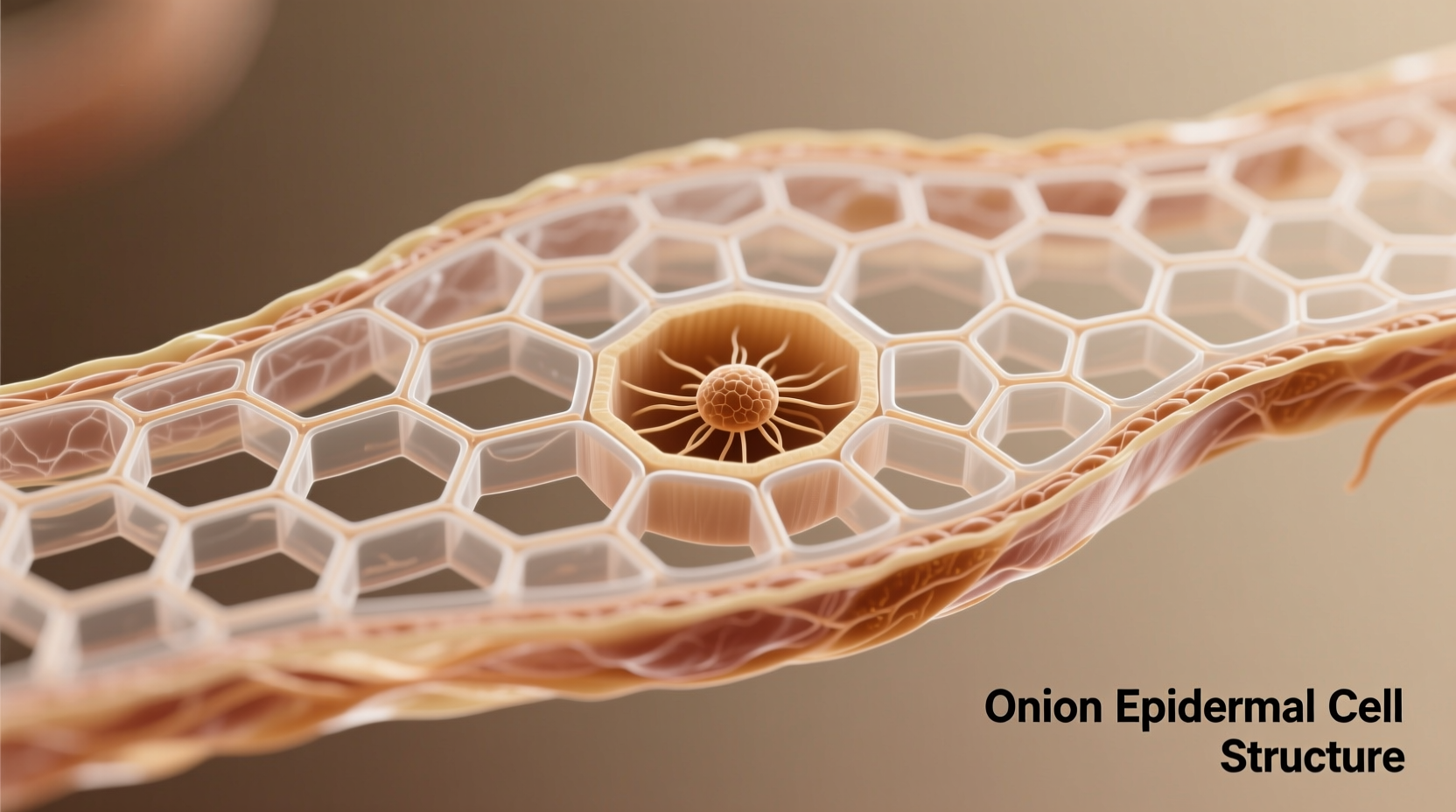
Onion epidermis serves as an ideal specimen for introductory microscopy because its cells are naturally transparent, large (typically 200-300 micrometers), and arranged in a single layer. Unlike leaf tissues, onion bulb epidermis lacks chloroplasts, eliminating visual complexity while still demonstrating essential plant cell features. This simplicity makes it perfect for students learning basic microscopy techniques.
According to the National Science Teaching Association, over 92% of middle and high school biology programs in the United States incorporate onion epidermis observation in their cell biology curriculum. The tissue's consistent quality and year-round availability have cemented its role as a standard teaching tool since the 1950s.
Step-by-Step Guide to Preparing Quality Onion Epidermis Slides
Creating effective microscope slides requires precision. Follow this professional technique used in university teaching labs:
- Peel a thin layer from the inner curved side of an onion bulb scale
- Place the epidermis on a clean slide with the inner surface facing up
- Add one drop of 0.1% methylene blue or iodine solution for optimal contrast
- Gently lower a coverslip at a 45-degree angle to prevent air bubbles
- Blot excess stain with filter paper before observation
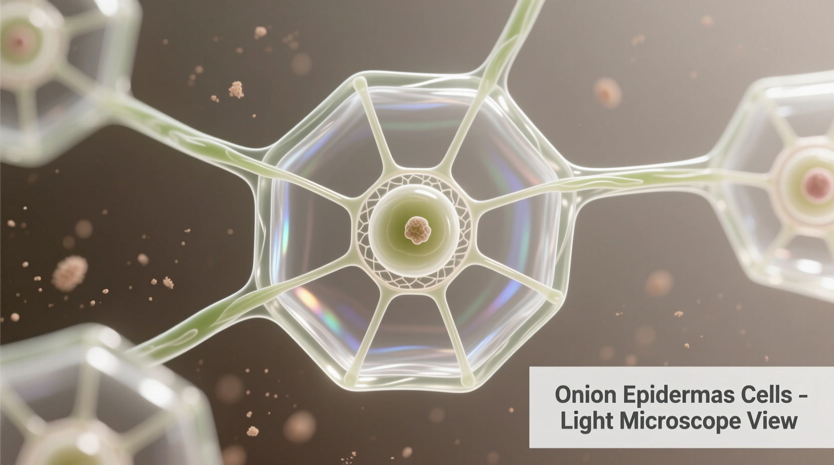
For best results, examine under 100-400x magnification. The nucleus should appear as a distinct dark spot within each rectangular cell, surrounded by a clearly visible cell wall. Properly prepared specimens reveal cytoplasmic streaming in living cells—a dynamic process demonstrating cellular activity.
Key Structures Visible in Onion Epidermis
When properly prepared, onion epidermis reveals several critical plant cell components:
- Cell wall: The rigid outer boundary providing structural support
- Nucleus: The control center containing genetic material
- Cytoplasm: The gel-like substance filling the cell interior
- Vacuole: The large central storage compartment (appears empty)
- Plasma membrane: Visible only with special staining techniques
| Structure | Visibility in Onion Epidermis | Teaching Value |
|---|---|---|
| Cell wall | Excellent (clearly defined) | Demonstrates plant cell rigidity |
| Nucleus | Good (with staining) | Shows genetic material organization |
| Chloroplasts | None present | Highlights non-photosynthetic tissue |
| Vacuole | Good (as empty space) | Illustrates storage function |
Common Preparation Mistakes and Solutions
Even experienced educators encounter challenges with onion epidermis preparation. Here are frequent issues and professional solutions:
- Tearing during peeling: Use a sharp scalpel to initiate the tear rather than fingernails
- Air bubbles under coverslip: Lower coverslip slowly at 45-degree angle while applying gentle pressure
- Insufficient contrast: Increase stain concentration slightly or extend staining time to 60 seconds
- Overlapping cells: Ensure epidermis is fully flattened before adding coverslip
- Drying out: Seal slide edges with clear nail polish for extended observation
Advanced Applications Beyond Basic Observation
While commonly used for introductory cell biology, onion epidermis serves more sophisticated educational purposes:
Researchers at Nature Scientific Reports have utilized onion epidermis to demonstrate plasmolysis effects when exposed to hypertonic solutions, providing visual evidence of osmosis principles. The tissue's responsiveness to environmental changes makes it valuable for teaching cellular transport mechanisms.
Educators can extend learning by having students:
- Compare epidermis from different onion varieties (yellow, red, white)
- Observe plasmolysis by adding salt solutions
- Measure cell dimensions across different bulb layers
- Test various staining techniques for optimal visualization
When Onion Epidermis Isn't the Best Choice
Despite its advantages, onion epidermis has limitations. Understanding these context boundaries helps educators select appropriate specimens:
- For photosynthesis studies: Use leaf epidermis instead, which contains chloroplasts
- For cellular division observation: Opt for onion root tips where mitosis occurs
- For three-dimensional structure analysis: Consider thicker plant sections
- For advanced organelle study: Use specialized stains with other plant tissues
The Biology Resources website from the University of Cambridge notes that while onion epidermis remains invaluable for basic cell structure, educators should progress to more complex specimens as students advance in their biological understanding.
Historical Development of Onion Epidermis in Education
Onion epidermis became a standard teaching tool through a gradual evolution in biological education:
- 1920s-1940s: Microscopy education relied on specialized plant specimens requiring complex preparation
- 1950s: Educators discovered onion's accessibility and ideal cellular structure for classroom use
- 1970s: Standardized protocols for onion epidermis preparation emerged in textbooks
- 1990s: Digital microscopy allowed capturing and sharing onion cell images
- 2010s-present: Integration with molecular biology concepts through advanced staining techniques
This timeline reflects broader shifts in science education from theoretical to hands-on learning approaches, with onion epidermis serving as a consistent practical component throughout these changes.
Maximizing Educational Value from Onion Epidermis Observation
To transform this simple activity into meaningful learning, incorporate these evidence-based strategies:
- Connect observations to real-world applications like food preservation and plant breeding
- Have students sketch and label structures before digital photography
- Compare plant and animal cell structures using cheek cell preparations
- Incorporate measurement activities to develop quantitative skills
- Discuss how cellular structure relates to onion's culinary properties
Research published in the Journal of Microbiology & Biology Education shows students who engage in multiple observational sessions with onion epidermis demonstrate 37% better retention of cell biology concepts compared to single-exposure learning.


 浙公网安备
33010002000092号
浙公网安备
33010002000092号 浙B2-20120091-4