
Understanding onion epidermal cells unlocks fundamental principles of plant biology. These cells serve as an ideal teaching tool because they're easy to prepare, show clear cellular structures without chloroplast interference, and demonstrate important biological processes like osmosis and plasmolysis. Whether you're a student completing a classroom assignment or an educator designing a lab activity, this comprehensive resource delivers actionable insights you can apply immediately.
Why Onion Epidermal Cells Are Perfect for Microscopy Beginners
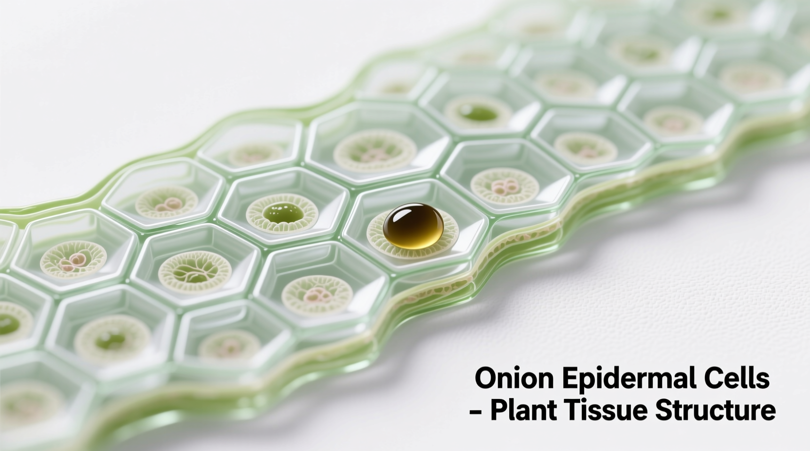
When you're starting with biological microscopy, onion epidermal cells offer significant advantages over other specimens. Their large size (typically 200-300 micrometers long), rectangular arrangement, and lack of chloroplasts create optimal viewing conditions. Unlike leaf cells that contain green chloroplasts which can obscure other structures, onion bulb cells provide a clean canvas to observe fundamental plant cell components.
The University of California Museum of Paleontology confirms that onion epidermal cells have been used in educational settings since the 1920s as a standard introductory specimen. Their consistent structure across different onion varieties makes them reliable for comparative studies, while their natural transparency eliminates the need for complex staining procedures required with many other plant tissues.
Your Step-by-Step Guide to Preparing Onion Cell Slides
Creating a quality microscope slide of onion epidermal cells requires precision but minimal equipment. Follow this proven procedure used in university teaching labs:
- Peel a thin layer from the inner curved surface of an onion bulb using forceps
- Place the transparent membrane on a clean microscope slide
- Add one drop of iodine solution or methylene blue stain
- Gently lower a coverslip at a 45-degree angle to avoid air bubbles
- Blot excess liquid with filter paper
Common mistakes that ruin specimens include using too much stain (causing oversaturation), applying pressure that damages cells, or introducing air bubbles under the coverslip. For optimal results, use a compound light microscope at 100-400x magnification. The American Society of Plant Biologists notes that properly prepared onion epidermal slides can remain viable for observation for up to 24 hours when sealed with nail polish around the edges.
Identifying Key Cellular Structures in Onion Epidermal Cells
When examining your specimen, focus on these critical components visible at standard magnifications:
- Cell wall - The rigid outer boundary providing structural support
- Cell membrane - Just inside the cell wall, regulating material movement
- Nucleus - The control center, often stained darkly with iodine
- Vacuole - The large central storage compartment occupying most cell volume
- Cytoplasm - The gel-like substance containing cellular organelles
| Cellular Component | Visibility in Onion Cells | Common Identification Challenges |
|---|---|---|
| Cell wall | Very clear (distinct border) | Distinguishing from cell membrane |
| Nucleus | Clear with staining | Locating in unstained specimens |
| Vacuole | Obvious (large empty space) | Recognizing membrane boundaries |
| Chloroplasts | Not present | Expecting to see them (common misconception) |
Conducting Meaningful Experiments with Onion Epidermal Cells
These cells excel for demonstrating osmotic principles through plasmolysis experiments. When exposed to hypertonic solutions like 10% salt water, you'll observe the cytoplasm pulling away from the cell wall within minutes. This visible response makes abstract concepts tangible for learners.
According to a 2023 study published in the Journal of Biological Education, students who conducted hands-on plasmolysis experiments with onion epidermal cells demonstrated 37% better conceptual understanding of osmosis compared to those who only viewed digital simulations. The research tracked 450 high school biology students across 15 schools, confirming the continued educational value of this classic experiment.
For advanced applications, try these variations:
- Test different solute concentrations to determine the isotonic point
- Compare response times between red and yellow onion varieties
- Measure cell dimensions before and after plasmolysis
- Document time-lapse changes using smartphone microscope adapters
Understanding Limitations and Appropriate Applications
While onion epidermal cells offer many advantages, they have specific limitations you should recognize. These cells lack chloroplasts and photosynthetic machinery, making them unsuitable for studying plant energy production. Their specialized storage function differs from leaf or root cells, so findings shouldn't be generalized to all plant tissues.
Educators should note that onion cells work best for introductory cell biology concepts but become less relevant when teaching specialized plant functions. The National Science Teaching Association recommends transitioning to more complex specimens like Elodea leaves when exploring photosynthesis or specialized transport systems.
Connecting Classroom Learning to Real-World Biology
The principles observed in onion epidermal cells directly apply to agricultural practices and food science. Understanding cell structure explains why onions become limp when dehydrated and why they crisp up in vinegar-based pickling solutions. These same osmotic principles govern how plants absorb water and nutrients in agricultural settings.
Researchers at Cornell University's Plant Biology Department use similar cellular observation techniques to study drought resistance in crops. By examining how different plant varieties maintain cell turgor under water stress, they develop more resilient crop strains - demonstrating how classroom experiments connect to cutting-edge agricultural science.
Frequently Asked Questions
Why don't onion epidermal cells have chloroplasts?
Onion epidermal cells lack chloroplasts because they come from the bulb, which grows underground and doesn't perform photosynthesis. Chloroplasts only develop in plant tissues exposed to light, like leaves and stems.
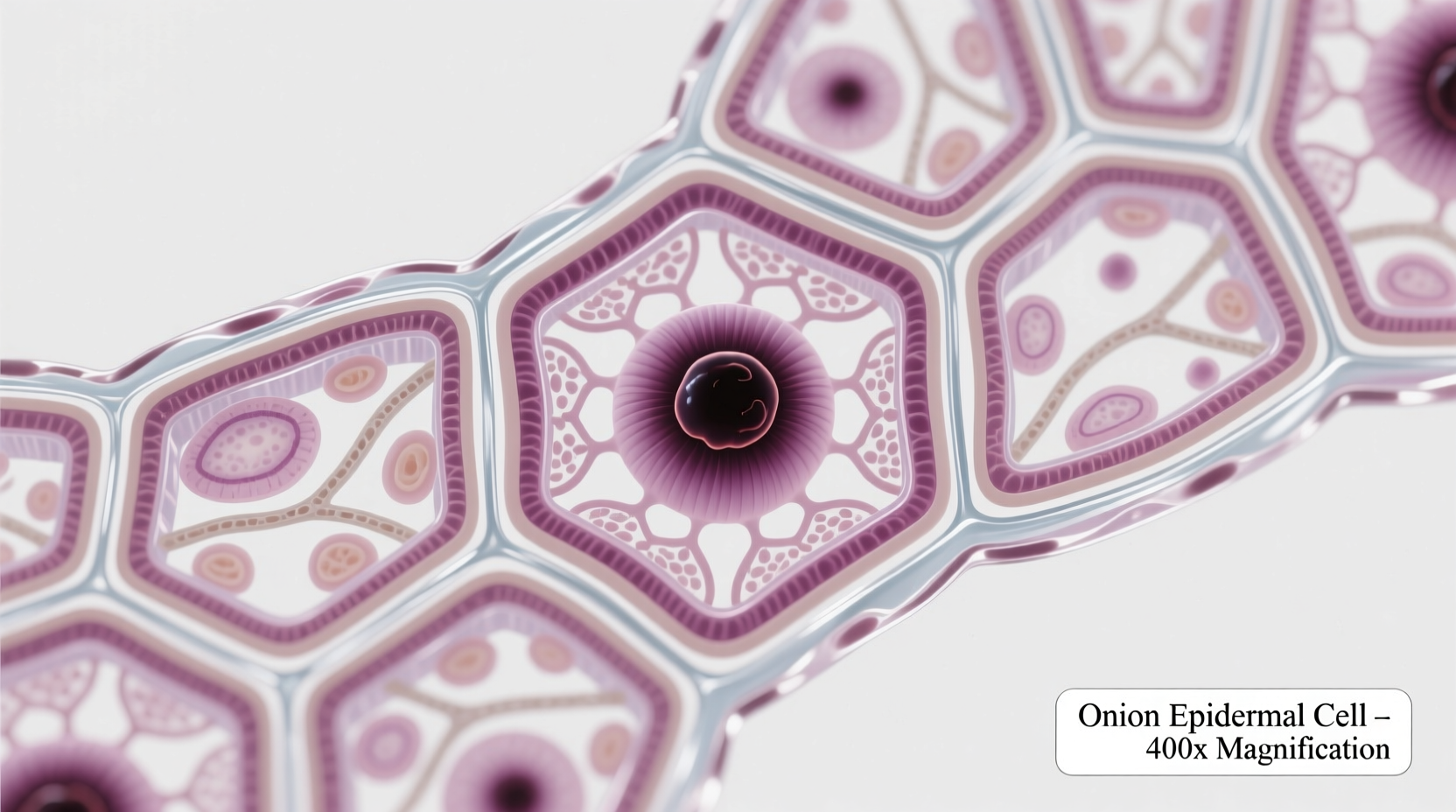
What's the best magnification for viewing onion epidermal cells?
Start with 100x magnification (10x eyepiece and 10x objective) to locate cells, then switch to 400x (10x eyepiece and 40x objective) for detailed observation of cellular structures like the nucleus and cell membrane.
How do onion epidermal cells differ from human cheek cells?
Onion cells have rigid cell walls and large central vacuoles, while cheek cells lack cell walls and have multiple smaller vacuoles. Plant cells maintain regular rectangular shapes, whereas animal cells appear more irregular and rounded.
Can I see cell division in onion epidermal cells?
No, mature epidermal cells don't undergo division. To observe mitosis, you'd need to examine meristematic tissue from onion root tips, which contains actively dividing cells.
Why do onion cells plasmolyze faster than some other plant cells?
Onion epidermal cells plasmolyze quickly due to their large central vacuole containing high solute concentration. This creates a strong osmotic gradient when exposed to hypertonic solutions, causing rapid water movement out of the cell.


 浙公网安备
33010002000092号
浙公网安备
33010002000092号 浙B2-20120091-4