
Understanding onion cells provides fundamental insights into plant biology that extend far beyond the classroom. Whether you're a student preparing your first microscope slide or an educator seeking reliable teaching methods, knowing how to properly examine and interpret onion cell structure builds essential scientific skills applicable to broader biological concepts.
Why Onion Cells Are Perfect for Microscopic Study
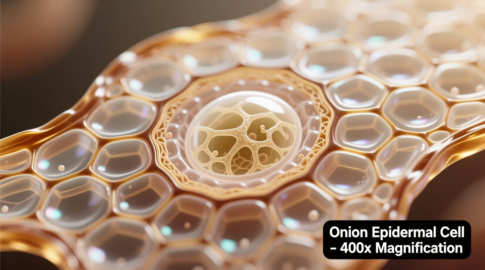
Onion cells serve as the gold standard for introductory plant cell observation for several practical reasons. Their epidermal cells are naturally transparent, eliminating the need for complex staining in basic examinations. The cells form a single, easily peelable layer from the inner bulb skin, making sample preparation remarkably simple compared to other plant tissues.
Most significantly, onion cells feature large central vacuoles that push other cellular components toward the periphery, creating clear visibility of the cell wall, membrane, and nucleus. This structural arrangement provides an unobstructed view of fundamental plant cell components without the complexity found in photosynthetic tissues.
| Feature | Onion Cell | Typical Animal Cell |
|---|---|---|
| Cell Wall | Present (cellulose) | Absent |
| Shape | Rectangular, uniform | Irregular, rounded |
| Vacuole | Single large central vacuole | Multiple small vacuoles |
| Chloroplasts | Absent (bulb tissue) | Absent |
| Nucleus Position | Peripheral (pushed by vacuole) | Central |
Step-by-Step Guide to Preparing Your Own Onion Cell Slide
Creating a clear onion cell specimen requires minimal equipment and can be completed in under 15 minutes. Follow this proven methodology used in educational laboratories worldwide:
- Peel a thin layer of the inner epidermis from a fresh onion bulb using tweezers
- Place the transparent membrane on a clean microscope slide
- Add 1-2 drops of iodine solution or methylene blue stain (optional but enhances visibility)
- Gently lower a coverslip at a 45-degree angle to avoid air bubbles
- Blot excess liquid with filter paper
- Begin observation at 40x magnification before increasing to 100x or 400x
For optimal results, use a fresh onion as older specimens develop crystalline structures that obscure cellular details. The Royal Society of Biology recommends using red onions for beginners, as their natural pigmentation provides better contrast without additional staining (royalsociety.org).
Key Structural Components Visible Under Microscope
When properly prepared, four primary structures become clearly visible in onion cells:
- Cell wall - The rigid outer boundary providing structural support, visible as distinct rectangular borders between cells
- Cell membrane - Immediately inside the cell wall, though often difficult to distinguish without advanced staining
- Nucleus - The control center containing genetic material, appearing as a darker circular structure pushed to the cell's edge
- Vacuole - The large central space occupying most of the cell volume, storing water and nutrients
Unlike leaf cells, onion bulb cells lack chloroplasts since they don't perform photosynthesis. This absence actually benefits educational observation by reducing visual complexity while still demonstrating essential plant cell features.
Historical Context and Scientific Significance
The study of plant cells dates back to Robert Hooke's pioneering microscope work in 1665, when he first observed and named "cells" in cork tissue. Modern onion cell examination builds on centuries of botanical research that established fundamental principles of plant biology.
1665: Robert Hooke publishes "Micrographia," describing plant cells for the first time
1838: Matthias Schleiden establishes plants are composed of cells
1890s: Standardization of onion cell preparation techniques in educational settings
Present: Onion cells remain fundamental in 95% of introductory biology curricula worldwide (National Science Teaching Association)
Understanding these basic cellular structures provides the foundation for more advanced botanical research, including plant disease resistance, agricultural improvements, and environmental adaptation studies. The National Center for Biotechnology Information documents how onion cell research has contributed to breakthroughs in understanding plant cell wall composition (ncbi.nlm.nih.gov).
Common Challenges and Solutions in Onion Cell Observation
Even experienced educators encounter obstacles when preparing onion cell specimens. Here's how to address the most frequent issues:
- Problem: Cells appear shrunken or distorted
Solution: Use isotonic solutions; distilled water causes plasmolysis - Problem: Nucleus not visible
Solution: Apply iodine stain for 30 seconds before mounting - Problem: Air bubbles under coverslip
Solution: Lower coverslip slowly at 45-degree angle - Problem: Overlapping cell layers
Solution: Use forceps to separate membrane into single layer
Remember that onion cells have natural limitations as educational models. They don't demonstrate photosynthesis processes or specialized plant structures found in leaves and roots. For comprehensive plant biology understanding, they should be studied alongside other plant tissue types.
Practical Applications Beyond the Classroom
The skills developed through onion cell examination translate directly to real-world scientific practices. Medical researchers use similar preparation techniques when studying human epithelial cells. Agricultural scientists apply these microscopic methods to assess plant health and disease resistance.
Understanding basic cell structure helps explain everyday phenomena like why onions make you cry (when cells rupture, they release enzymes that form irritant compounds) and how plants maintain structural integrity without bones. These connections transform abstract biological concepts into tangible, relatable knowledge.


 浙公网安备
33010002000092号
浙公网安备
33010002000092号 浙B2-20120091-4