
Discovering what an onion looks like under a microscope reveals the fundamental building blocks of plant life. This simple yet profound observation forms the cornerstone of biology education worldwide, helping students visualize cellular structures that are otherwise invisible to the naked eye. Whether you're a student completing a science project, a teacher preparing a classroom demonstration, or simply curious about the microscopic world, understanding onion cell structure provides valuable insights into plant biology.
Why Onion Cells Make the Perfect Microscopic Subject
Onion epidermis serves as an ideal specimen for microscopy beginners for several practical reasons. The outer skin layers are thin enough to transmit light effectively, require minimal preparation, and display clear cellular structures even at low magnification. Unlike many plant tissues, onion cells don't contain chloroplasts, making the nucleus and other organelles more visible without color interference. This characteristic makes onion cells particularly valuable for educational settings where students are learning basic microscopy techniques.
What You'll Actually See at Different Magnifications
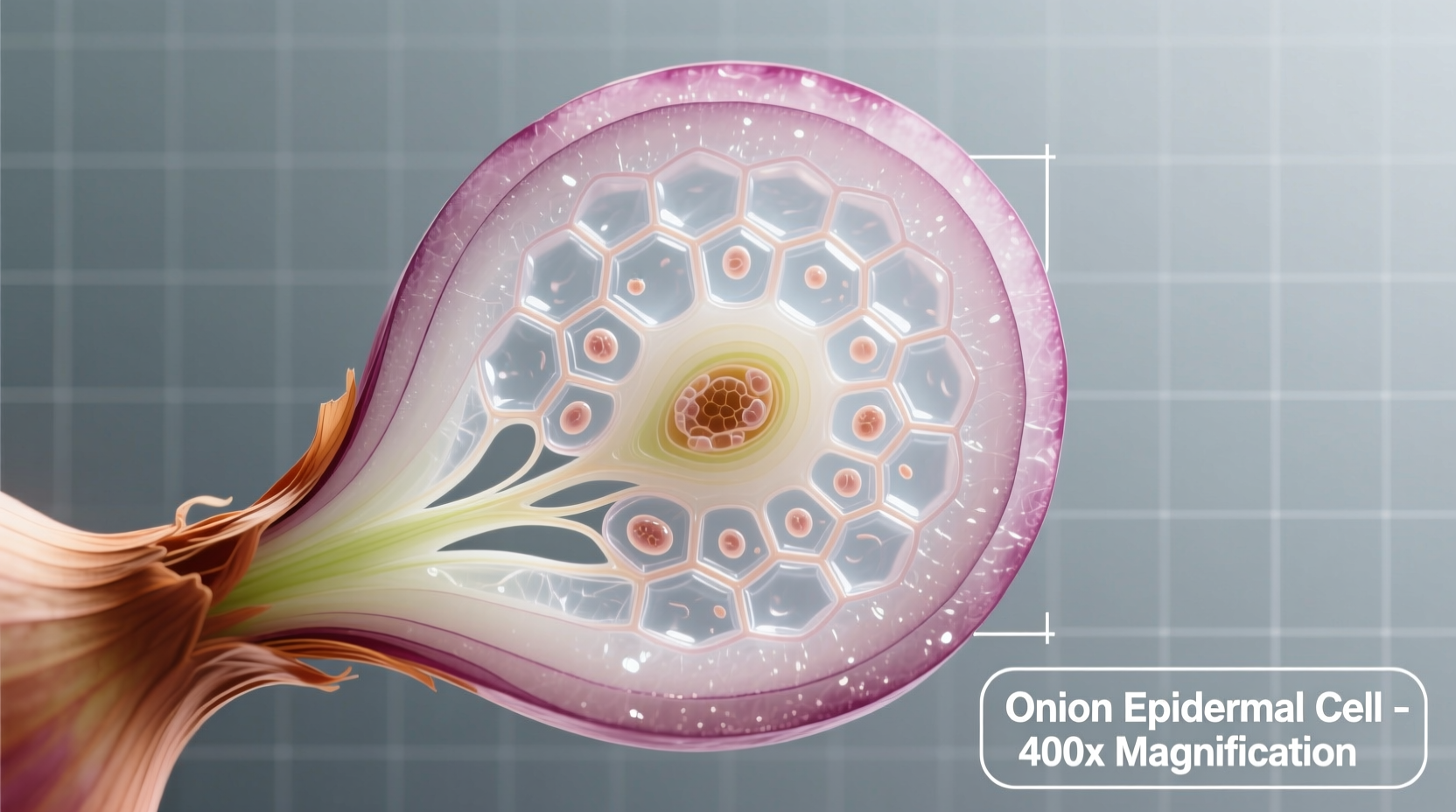
Understanding the onion under microscope experience requires knowing what to expect at various magnification levels. At 40x magnification, you'll see the overall arrangement of cells in a brick-like pattern. Increase to 100x, and cell walls become distinctly visible with occasional nuclei appearing as darker spots. At 400x magnification—the maximum for standard light microscopes—you can observe detailed cellular components including the nucleus, cytoplasm, and the large central vacuole that characterizes plant cells.
| Magnification Level | Visible Structures | Best For |
|---|---|---|
| 40x | Overall cell arrangement, basic rectangular pattern | Initial observation, locating area of interest |
| 100x | Clear cell walls, visible nuclei, cytoplasm boundaries | Detailed plant cell structure observation |
| 400x | Nucleus details, possible organelles, cell membrane | Advanced cellular component study |
Preparing Your Own Onion Slide: A Step-by-Step Guide
Creating a quality onion slide requires minimal equipment but precise technique. Start by carefully peeling a thin layer of the inner epidermis from a fresh onion bulb—aim for a piece about the size of a postage stamp. Place this specimen on a clean microscope slide and add one drop of iodine solution or methylene blue stain to enhance visibility of cellular structures. Gently lower a coverslip at a 45-degree angle to avoid air bubbles, which can obstruct your view. This simple onion slide preparation technique works effectively for classroom demonstrations and home experiments alike.
Many beginners struggle with common issues like air bubbles or uneven staining. If your cells appear distorted, you've likely applied too much pressure when placing the coverslip. For clearer nuclei visibility, allow the stain to sit for 30-60 seconds before viewing. Remember that fresh onion specimens yield the best results—older onions may show degraded cellular structures that make observation difficult.
Scientific Significance of Onion Cell Structure
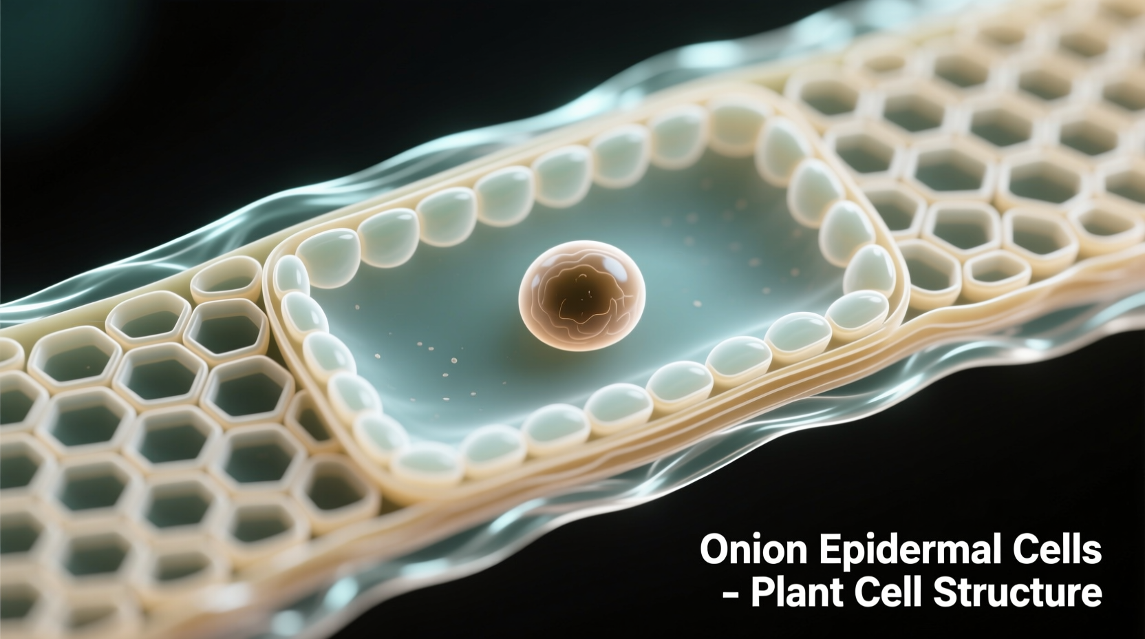
The rectangular arrangement of onion cells under microscope reveals fundamental principles of plant biology. Unlike animal cells, plant cells maintain their shape through rigid cellulose cell walls, creating the distinctive grid pattern visible even at low magnification. The large central vacuole, which occupies approximately 80% of the cell's volume, stores water and maintains turgor pressure essential for plant structure. The nucleus, typically visible as a darker circular structure near the cell wall, contains the genetic material that directs cellular activities.
Historically, onion cells have played a crucial role in biological education since the development of light microscopy in the 19th century. According to educational resources from the National Science Teaching Association, onion epidermis became a standard teaching specimen because of its consistent cellular structure and ease of preparation. This historical context explains why "onion under microscope" remains one of the most common introductory biology experiments worldwide.
Troubleshooting Common Microscopy Challenges
When viewing onion cells under microscope, several issues commonly arise. If cells appear transparent with no visible structures, insufficient staining is likely the problem—try increasing stain concentration or duration. Blurry images typically indicate improper focus or dirty lenses; clean your microscope optics with appropriate lens paper. For specimens where cell walls appear broken or irregular, you've probably damaged the delicate epidermis during preparation.
Understanding these context boundaries helps set realistic expectations. Onion cells from different varieties may show slight variations in size and shape, but the fundamental structure remains consistent. Environmental factors like storage conditions can affect cellular integrity—freshly prepared specimens always yield superior results. Remember that light microscope limitations mean you won't see organelles like mitochondria or ribosomes; these require electron microscopy for visualization.


 浙公网安备
33010002000092号
浙公网安备
33010002000092号 浙B2-20120091-4