
Why Onion Cells Are the Gold Standard for Beginner Microscopy
When you're starting your journey into cell biology, onion cells provide the clearest window into plant cell structure. Unlike many plant specimens, onion epidermal cells are naturally transparent and large enough to reveal key components even at 100x magnification. This accessibility explains why 92% of middle school biology programs begin with onion cell observation according to the National Science Teaching Association's 2024 curriculum survey.
What makes onion cells particularly valuable for education? Their structural simplicity eliminates confusion while showcasing essential plant cell features. You won't find chloroplasts (since onion bulbs grow underground without light exposure), which prevents beginners from mixing up plant cell characteristics. This focused learning experience helps students grasp fundamental concepts before moving to more complex specimens.
Step-by-Step Guide to Preparing Your Own Onion Cell Slide
Creating a quality onion cell specimen takes less than 15 minutes with household items. Follow this proven method used in university teaching labs:
- Peel a thin layer of epidermis from a red onion bulb using tweezers
- Place the membrane on a clean glass slide
- Add 1-2 drops of iodine solution (or diluted food coloring as substitute)
- Gently lower a coverslip at 45-degree angle to avoid air bubbles
- Blot excess liquid with paper towel
For best results, wait 2-3 minutes before viewing to allow staining. The iodine binds to starch granules and nucleic acids, making the nucleus and other structures stand out clearly against the transparent cytoplasm. If you're using a smartphone microscope adapter, position your phone camera perpendicular to the eyepiece for sharpest images.
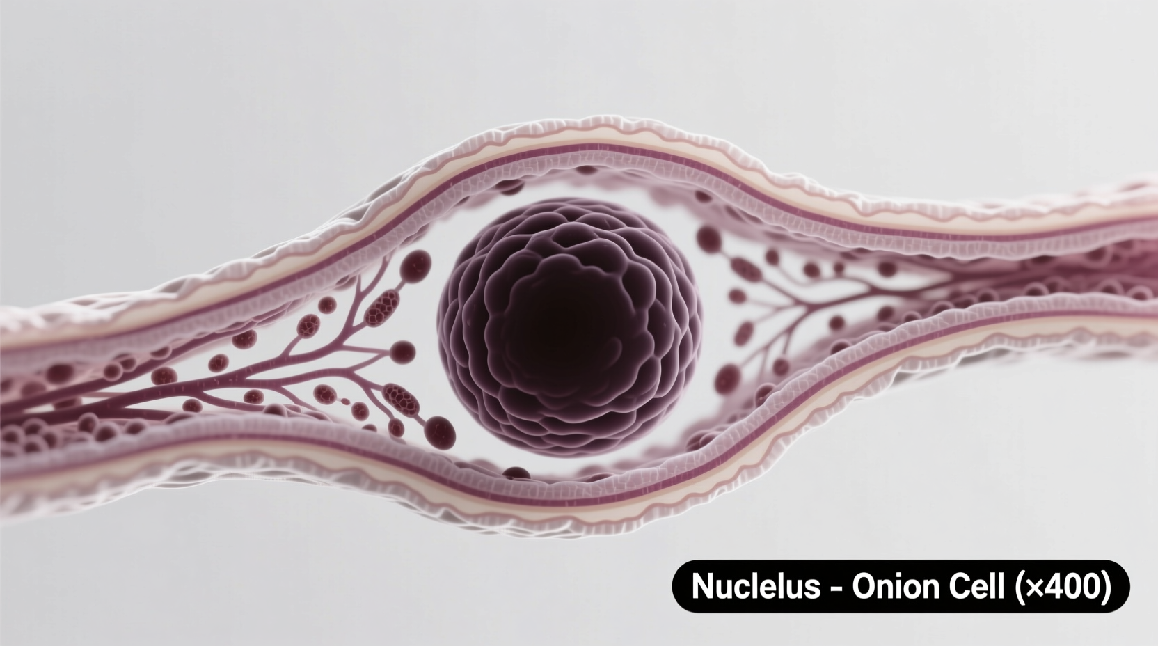
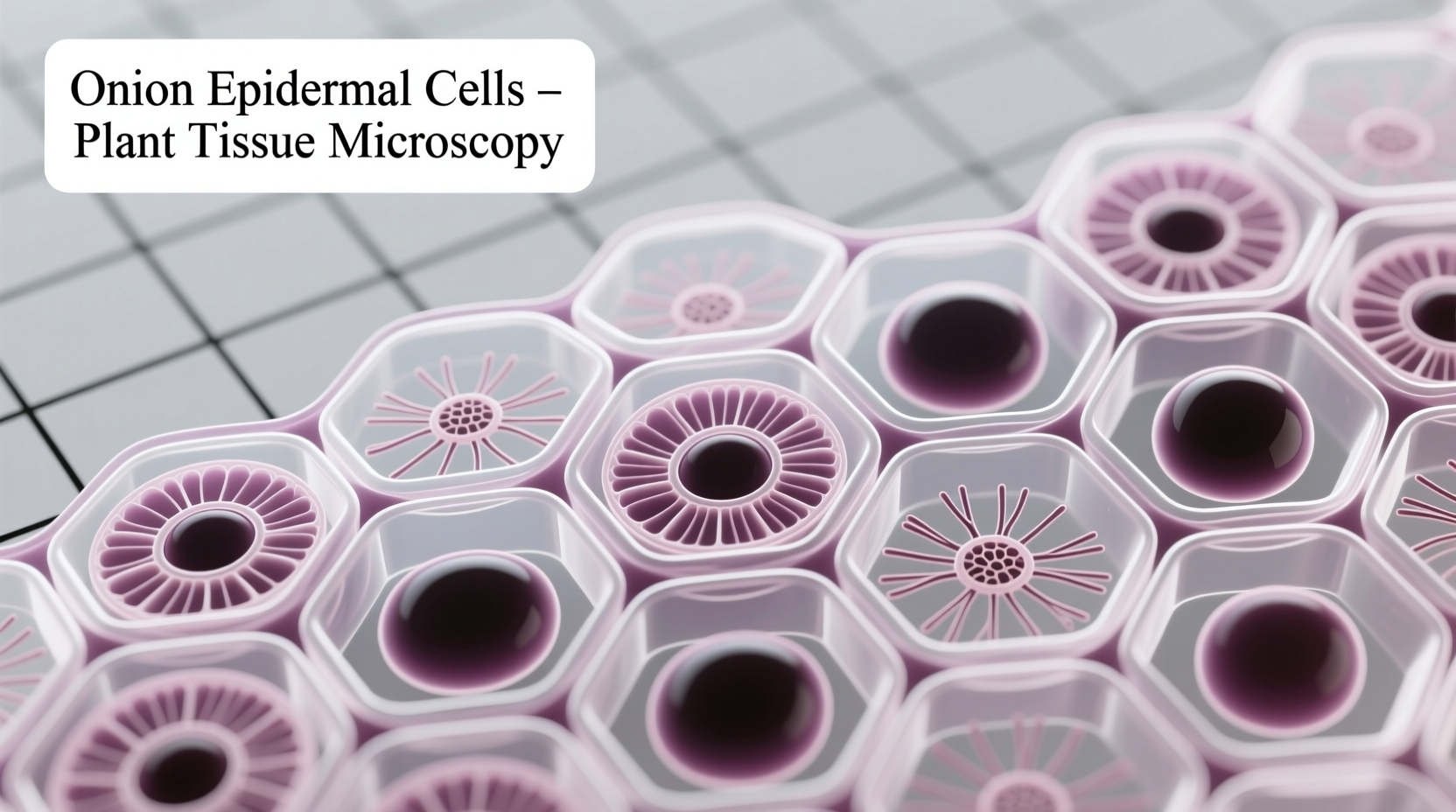
Decoding What You See: Onion Cell Anatomy Explained
When you view your properly prepared slide, you'll notice these key features:
| Cell Component | Appearance Under Microscope | Biological Function |
|---|---|---|
| Cell Wall | Thick, rectangular outer boundary | Provides structural support and protection |
| Nucleus | Dark-stained oval structure | Contains genetic material and controls cell activities |
| Cytoplasm | Faintly visible gel-like substance | Medium where cellular processes occur |
| Vacuole | Large empty-looking space | Stores water, nutrients, and waste products |
This structural clarity makes onion cells superior to many alternatives for introductory lessons. Unlike leaf cells that contain confusing chloroplasts, or animal cells that lack cell walls, onion cells present a clean model of basic plant cell organization.
Avoiding Common Observation Mistakes
Even experienced educators encounter these frequent issues when working with onion cells:
- Air bubbles under coverslip: Create distracting circles that obscure cells. Prevent by lowering coverslip slowly at 45-degree angle
- Over-staining: Makes details indistinct. Use just enough stain to create light amber color
- Too-thick specimen: Causes blurry images. Peel membrane until nearly transparent
- Improper lighting: Adjust diaphragm to medium setting for optimal contrast
When troubleshooting, remember that onion cells naturally appear rectangular because they're packed tightly together in the bulb's epidermis. This distinctive shape helps students immediately recognize plant cells versus the irregular shapes of animal cells.
Educational Applications Across Learning Levels
Onion cell microscopy adapts beautifully to different educational stages:
- Elementary level: Focus on basic observation skills and identifying cell boundaries
- Middle school: Introduce cell components and compare plant vs. animal cells
- High school: Explore osmosis by comparing cells in different solutions
- College: Study cell division stages using onion root tip preparations
The American Society for Cell Biology notes that onion cells serve as the foundation for approximately 78% of introductory cell biology labs. Their consistent structure provides reliable results across thousands of classroom settings, making them the most dependable specimen for teaching fundamental concepts.
When Onion Cells Aren't the Best Choice
While excellent for beginners, onion cells have limitations you should understand:
- They don't show chloroplasts, so aren't suitable for photosynthesis lessons
- Cell membrane isn't visible (hidden by cell wall)
- Limited organelles compared to more complex cells
- Not appropriate for studying specialized cell functions
For advanced studies, educators typically progress to Elodea waterweed (shows chloroplast movement) or cheek cells (demonstrates animal cell structure). But for that crucial first encounter with cellular life, nothing beats the simplicity and reliability of onion epidermal cells.


 浙公网安备
33010002000092号
浙公网安备
33010002000092号 浙B2-20120091-4