
Why Onion Cells Are Perfect for Microscopy Beginners
When you examine an onion cell under a microscope, you're observing one of the most accessible plant cell models available. The epidermal layer of onion skin provides a single-cell-thick specimen that's naturally translucent, requiring minimal preparation to reveal key cellular structures. Unlike many plant tissues, onion cells don't contain chloroplasts, eliminating green pigment interference and allowing clearer visualization of the nucleus and other organelles.
For students and educators, this experiment delivers immediate results with basic equipment. Within 10 minutes, you can prepare a slide showing definitive plant cell characteristics at 100x magnification. The distinctive rectangular shape created by rigid cell walls makes identification straightforward, even for microscopy beginners. This practical approach transforms abstract cell theory into tangible learning.
Step-by-Step Guide: Preparing Your Onion Cell Slide
Follow this proven procedure to create a high-quality onion cell specimen that reveals maximum detail:
Materials You'll Need
- Fresh onion bulb (white or yellow varieties work best)
- Microscope glass slides and coverslips
- Distilled water
- Iodine solution ( Lugol's iodine) or methylene blue stain
- Forceps and scalpel or razor blade
- Dropper
- Blotting paper or paper towels
Preparation Process
- Peel a thin layer of the inner epidermis from a fresh onion bulb using forceps
- Place the transparent membrane on a clean glass slide
- Add 1-2 drops of distilled water to prevent drying
- Gently lower a coverslip at a 45-degree angle to avoid air bubbles
- For enhanced visibility, add a drop of iodine solution at one edge of the coverslip and draw it through with blotting paper
- Allow 30 seconds for staining, then view under microscope
| Common Problem | Why It Happens | Solution |
|---|---|---|
| Overlapping cells | Membrane too thick | Use forceps to separate layers further |
| Fuzzy appearance | Air bubbles under coverslip | Lower coverslip more slowly at angle |
| Poor nucleus visibility | Insufficient staining time | Wait 45-60 seconds before viewing |
| Drying specimen | Water evaporating | Seal edges with petroleum jelly |
What You'll See: Onion Cell Anatomy Explained
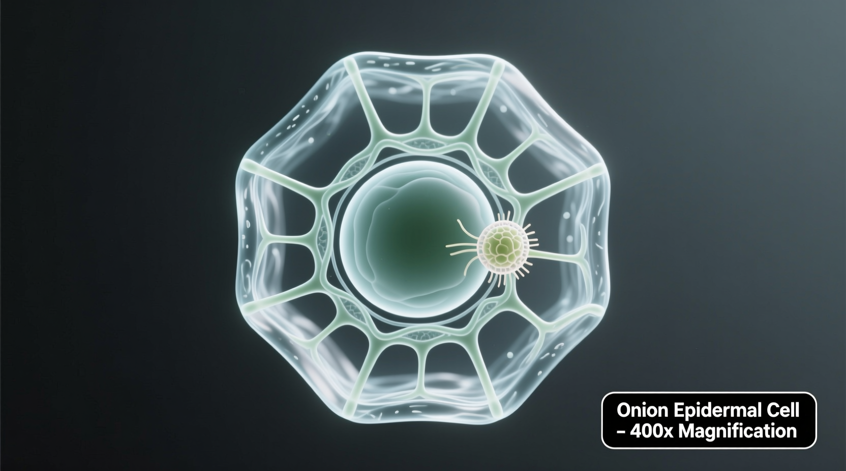
At 100x magnification, onion cells display their characteristic brick-like arrangement. The most prominent feature is the rigid cell wall composed of cellulose, giving plant cells their distinctive rectangular shape. Inside this wall, you'll observe the cell membrane (though less visible without special staining), cytoplasm appearing as a semi-transparent matrix, and the nucleus stained dark brown by iodine solution.
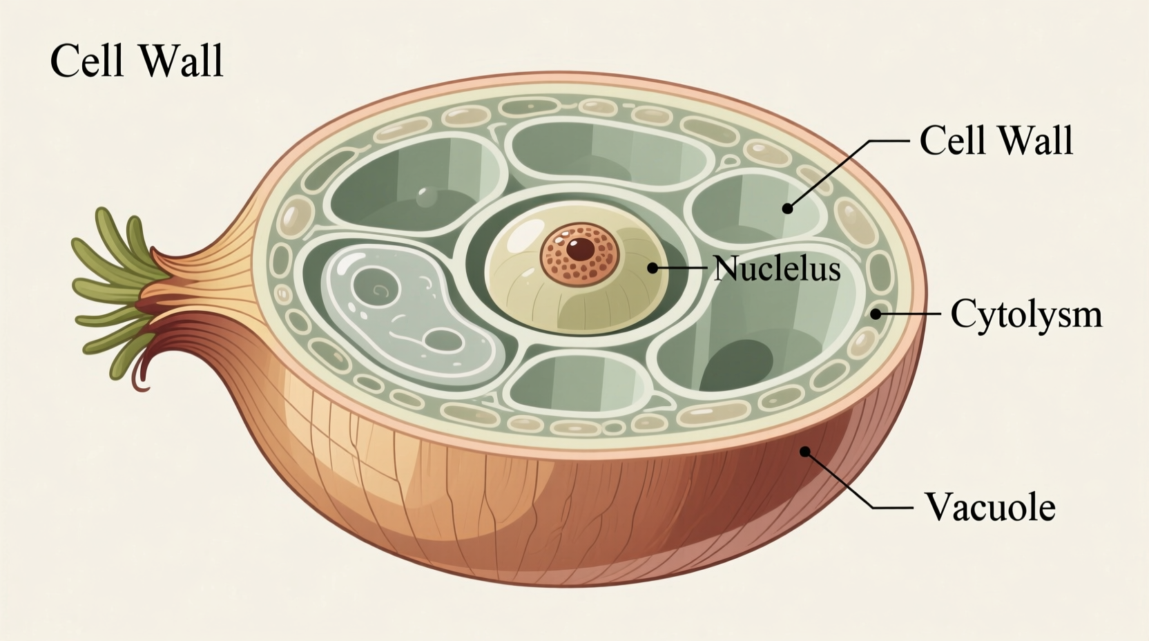
Unlike animal cells, onion cells lack centrioles and have a large central vacuole that occupies most of the cell volume. This vacuole contains cell sap and helps maintain turgor pressure. The absence of chloroplasts in onion epidermal cells makes them particularly valuable for studying basic cell structures without pigment interference.
Historical Context: Milestones in Plant Cell Observation
The study of plant cells through microscopy has evolved significantly since Robert Hooke's pioneering work. Understanding this timeline helps appreciate why onion cells remain a staple in biology education:
| Year | Scientific Advancement | Educational Impact |
|---|---|---|
| 1665 | Robert Hooke observes cork cells | Coins term "cell" from monastery rooms |
| 1838 | Schleiden identifies plant cells as basic units | Establishes cell theory foundation |
| 1940s | Electron microscopy reveals organelles | Confirms onion cell structures seen with light microscopes |
| Present | Digital microscopy accessible to classrooms | Students capture and analyze onion cell images digitally |
Practical Applications and Limitations
While onion cells provide an excellent introduction to plant cell biology, understanding their context boundaries enhances experimental value. These cells work exceptionally well for demonstrating basic plant cell structures but have specific limitations educators should recognize.
Onion epidermal cells excel at showing cell walls, nuclei, and cytoplasm organization, making them perfect for introductory biology. However, they don't display chloroplasts since they come from underground bulb tissue, limiting photosynthesis-related studies. For observing active cellular processes like mitosis, root tip cells provide better specimens.
According to the National Science Teaching Association's laboratory guidelines, onion cell preparation remains one of the top recommended activities for middle and high school biology because it requires minimal equipment while delivering clear educational outcomes. The American Society of Plant Biologists confirms that this experiment effectively demonstrates fundamental plant cell characteristics applicable to understanding more complex plant tissues.
Advanced Techniques for Enhanced Observation
Once you've mastered basic onion cell preparation, these advanced methods will reveal additional cellular details:
Plasmolysis Demonstration
Create a dramatic demonstration of osmosis by adding 10% salt solution to your slide. Within minutes, you'll observe the cytoplasm pulling away from the cell wall as water exits the cell. This visible process demonstrates how plant cells respond to hypertonic environments.
Fluorescent Staining Options
For more detailed nuclear observation, substitute iodine with aceto-orcein stain. This specialized stain binds to DNA, making chromosome structures visible during cell division. While requiring slightly more advanced preparation, this technique reveals cellular processes beyond basic structure.
Digital Image Analysis
Capture images of your onion cells using a smartphone adapter. Free software like ImageJ allows measurement of cell dimensions and calculation of magnification. Students can create comparative charts of cell size across different onion varieties or growth conditions.
Troubleshooting Common Observation Problems
Even experienced educators encounter challenges with onion cell microscopy. Here's how to address frequent issues:
- Nucleus not visible: Increase staining time to 60 seconds and ensure proper iodine concentration (1-2%)
- Overlapping cells: Use younger onion bulbs and separate membrane layers more carefully
- Cloudy appearance: Clean slides thoroughly with alcohol before use
- Rapid drying: Seal slide edges with petroleum jelly or use commercial mounting medium
- Poor contrast: Adjust microscope diaphragm to optimize light conditions
Remember that onion cell quality varies by season. Spring-harvested onions typically yield better specimens than older, stored bulbs. For consistent classroom results, purchase fresh onions weekly during lab activities.


 浙公网安备
33010002000092号
浙公网安备
33010002000092号 浙B2-20120091-4