
Why Onion Bulb Cells Matter in Biological Studies
When you examine onion bulb cells under a microscope, you're observing one of the most accessible windows into plant cell biology. These cells serve as the perfect educational model because they're large (typically 200-300 micrometers), easy to prepare, and display clear structural components without complex pigmentation. Unlike leaf cells, onion bulb cells lack chloroplasts since they grow underground away from light, creating a clean canvas for identifying fundamental cellular structures.
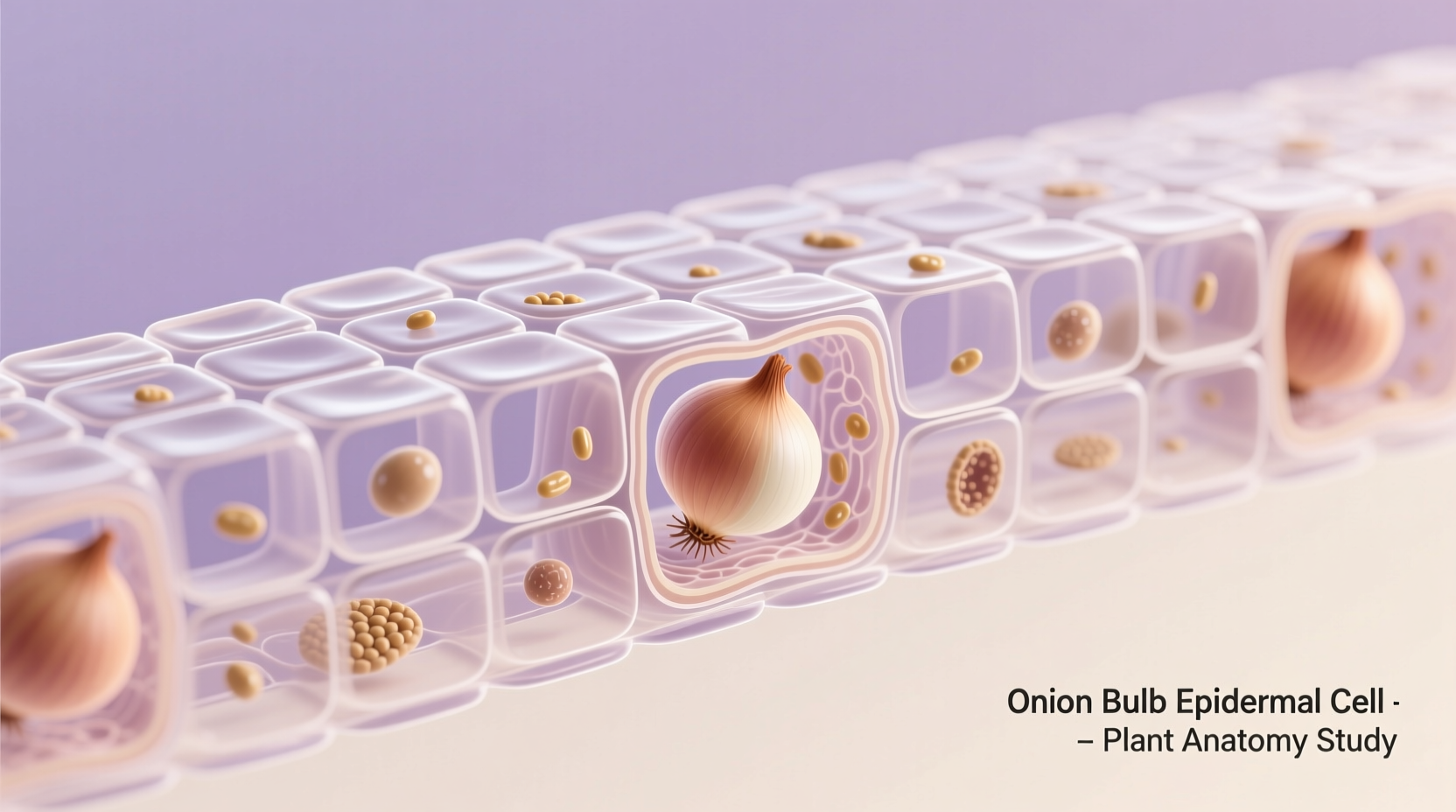
For students beginning their journey in cell biology, onion bulb cells offer immediate visual recognition of key components. The distinct cell wall provides structural integrity while the large central vacuole occupies approximately 90% of the cell's volume, pushing other organelles toward the periphery. This arrangement makes the nucleus particularly visible when stained with iodine or methylene blue solutions.
Anatomy of an Onion Bulb Cell: Key Components
Understanding the specific structures within onion bulb cells helps build foundational knowledge for more complex biological concepts. Each component serves essential functions that maintain cellular integrity and facilitate biological processes.
| Cellular Component | Appearance Under Microscope | Primary Function |
|---|---|---|
| Cell Wall | Clear rectangular boundary | Provides structural support and protection |
| Cell Membrane | Thin line just inside cell wall | Regulates substance movement in/out of cell |
| Nucleus | Dark spot when stained | Contains genetic material and controls cell activities |
| Cytoplasm | Faint granular area surrounding nucleus | Medium for cellular processes and organelle suspension |
| Vacuole | Large empty-looking space | Stores water, nutrients, and maintains turgor pressure |
Practical Guide to Observing Onion Bulb Cells
Preparing your own onion bulb cell slide takes just minutes with basic equipment. Follow these steps for clear microscopic viewing:
- Peel a thin layer of epidermis from the inner curved side of an onion bulb
- Place the tissue on a clean microscope slide
- Add 1-2 drops of iodine or methylene blue stain
- Cover with a coverslip using the water-mount technique
- Gently press to spread the cells without damaging them
- View under 100x magnification first, then increase to 400x
When properly prepared, you'll immediately notice the distinctive brick-like arrangement of cells. The staining process enhances visibility of the nucleus, which appears as a dark spot near the cell wall. This hands-on experience helps solidify theoretical knowledge through direct observation.

Historical Context: Onion Cells in Science Education
The use of onion bulb cells in educational settings spans more than a century, evolving alongside microscopy technology. This timeline reveals how these simple cells became fundamental teaching tools:
| Time Period | Development | Educational Impact |
|---|---|---|
| 1830s-1850s | Early compound microscopes become widely available | Scientists first document plant cell structures using onion specimens |
| 1880s | Standardization of biological teaching materials | Onion cells become standard curriculum component in European universities |
| 1920s | Introduction of affordable student microscopes | Onion cell experiments enter high school biology classrooms |
| 1950s-present | Digital microscopy and enhanced staining techniques | Modern classrooms use onion cells to teach cell theory and microscopy skills |
Common Misconceptions About Onion Bulb Cells
Several misunderstandings persist about onion bulb cells that can confuse learners. Let's clarify these points with scientific accuracy:
- Myth: Onion bulb cells contain chloroplasts
Fact: Since bulb cells develop underground without light exposure, they lack chloroplasts. Only the green leaf portions of onions contain these photosynthetic organelles. - Myth: The large empty space is an air pocket
Fact: This is actually the central vacuole filled with cell sap, crucial for maintaining turgor pressure and storing nutrients. - Myth: All plant cells look identical to onion bulb cells
Fact: Plant cells vary significantly by tissue type and species. Onion bulb cells represent just one specialized form adapted for storage.
Scientific Significance Beyond the Classroom
While primarily known for educational purposes, onion bulb cells contribute to meaningful scientific research. Their transparent nature makes them valuable for studying cellular responses to environmental stressors. Researchers at institutions like the University of California, Davis have used onion cells to investigate:
- Effects of osmotic pressure changes on cell structure
- Cellular responses to pollutants and toxins
- Basic mechanisms of plasmolysis and deplasmolysis
- Early indicators of cellular damage from radiation
These studies provide foundational knowledge applicable to agricultural science and environmental monitoring. The simplicity of onion bulb cells allows researchers to isolate specific cellular processes without complex variables.
Comparing Onion Bulb Cells to Other Biological Specimens
Understanding how onion bulb cells differ from other common microscopy specimens helps contextualize their educational value. Each specimen offers unique advantages for studying specific cellular features:
| Specimen Type | Key Advantages | Limitations | Best For Studying |
|---|---|---|---|
| Onion Bulb Cells | Large size, clear structure, easy preparation | No chloroplasts, limited organelle variety | Basic plant cell structure, cell wall function |
| Elodea Leaf Cells | Contains active chloroplasts, visible cytoplasmic streaming | Smaller size, more complex structure | Photosynthesis, organelle movement |
| Human Cheek Cells | Represents animal cell structure, easy collection | Small size, fragile membrane, no cell wall | Animal cell comparison, nucleus structure |
| Potato Cells | Contains starch granules, different storage function | Darker appearance, less distinct boundaries | Storage mechanisms, starch identification |
Advanced Applications in Modern Biology
Recent technological advances have expanded the utility of onion bulb cells beyond basic education. Researchers now use these cells for more sophisticated investigations:
At the cellular level, scientists employ onion bulb cells to study programmed cell death (apoptosis) mechanisms. The transparent nature of these cells allows researchers to observe cellular changes during stress responses without complex imaging techniques. According to research published in the Journal of Plant Physiology, onion cells demonstrate clear morphological changes during osmotic stress that mirror responses in more complex crop plants.
For environmental science applications, educators and researchers use onion root tip cells (a related specimen) to study mitosis and chromosome behavior. While technically different from bulb cells, this application demonstrates how onion tissue specimens contribute to understanding fundamental biological processes.
Practical Tips for Successful Microscopy Sessions
Whether you're a student or educator, these evidence-based techniques will improve your onion bulb cell observations:
- Peel selection: Choose the innermost layers of the bulb for the thinnest, most transparent specimens
- Staining technique: Use iodine solution for 30-60 seconds then gently blot excess to prevent over-staining
- Focusing strategy: Start with lowest magnification to locate cells, then gradually increase
- Light adjustment: Reduce light intensity when viewing at higher magnifications for better contrast
- Measurement practice: Use stage micrometers to calculate actual cell dimensions
These practical approaches address common challenges students face when first examining biological specimens. Proper technique ensures clear visualization of cellular components, transforming abstract concepts into tangible observations.
Frequently Asked Questions
Why don't onion bulb cells have chloroplasts?
Onion bulb cells develop underground where they receive no light exposure. Since chloroplasts require light for photosynthesis, these storage cells evolved without them. Only the green leaf portions of the onion plant contain chloroplasts, as they're exposed to sunlight.
What causes the rectangular shape of onion bulb cells?
The rectangular shape results from the rigid cellulose cell wall that maintains structural integrity. Unlike animal cells with flexible membranes, plant cells pack tightly together in organized layers, creating the characteristic brick-like pattern visible under magnification.
How do you measure the size of onion bulb cells accurately?
To measure onion bulb cells, first calibrate your microscope using a stage micrometer. Then, view your specimen at 400x magnification and use an ocular micrometer to measure cell dimensions. Typical onion bulb cells range from 200-300 micrometers in length, with width approximately half that measurement.
Why does the nucleus appear in different positions in various cells?
The nucleus position varies because the large central vacuole pushes cellular components toward the periphery. In some cells, the nucleus appears near the cell wall, while in others it might be more central, depending on the cell's developmental stage and environmental conditions.
Can you observe cell division in onion bulb cells?
While possible, onion bulb cells aren't ideal for observing cell division. The actively dividing cells are found in onion root tips, not the bulb tissue. Root tip preparations provide clearer examples of mitosis stages, which is why they're commonly used for studying cell division processes.


 浙公网安备
33010002000092号
浙公网安备
33010002000092号 浙B2-20120091-4