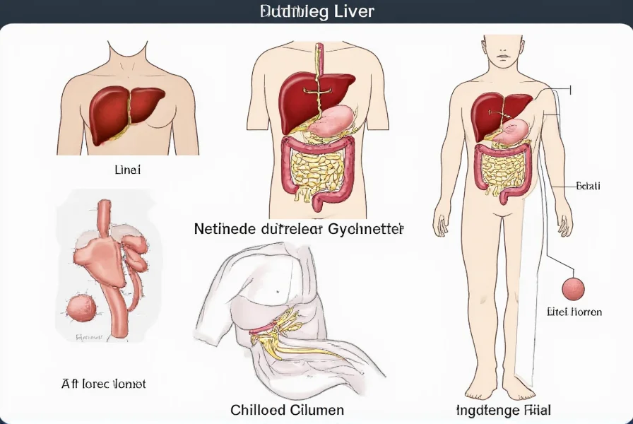
Nutmeg liver represents a classic histopathological finding in chronic passive congestion of the liver. This condition develops when blood backs up in the hepatic veins due to impaired venous return, most commonly from right heart failure. The name derives from the visual resemblance to a nutmeg spice surface, with dark red congested centrilobular areas surrounded by paler periportal regions.
Understanding Nutmeg Liver Pathology
The development of nutmeg liver follows a specific pathological progression. When venous pressure increases in the hepatic veins, blood pools in the centrilobular regions (zone 3) of the liver lobule. This congestion leads to hypoxia, hepatocyte atrophy, and eventually centrilobular necrosis. Meanwhile, the periportal regions (zone 1) maintain relatively better oxygenation, creating the characteristic alternating pattern.
Primary Causes of Nutmeg Liver
Chronic venous congestion leading to nutmeg liver typically stems from conditions that impair right heart function or obstruct hepatic venous outflow:
| Cause Category | Specific Conditions | Frequency |
|---|---|---|
| Cardiac Disorders | Congestive heart failure, tricuspid regurgitation, constrictive pericarditis | Most common (85-90%) |
| Hepatic Vein Obstruction | Budd-Chiari syndrome, hepatic vein thrombosis | 5-10% |
| Inferior Vena Cava Issues | IVC obstruction, stenosis, or thrombosis | 2-5% |
Clinical Presentation and Symptoms
Patients with nutmeg liver typically present with symptoms related to the underlying cause rather than the liver appearance itself. Common manifestations include:
- Right upper quadrant abdominal discomfort or fullness
- Hepatomegaly (enlarged liver) that's often tender
- Ascites in advanced cases
- Peripheral edema
- Jugular venous distension
- Progressive fatigue and exercise intolerance
It's important to note that nutmeg liver itself isn't a disease but rather a pathological finding indicating chronic venous congestion. Many patients remain asymptomatic until significant cardiac compromise occurs.
Diagnostic Approaches for Nutmeg Liver
Diagnosing the underlying condition causing nutmeg liver involves multiple approaches:
Imaging Studies
Ultrasound with Doppler is typically the first-line imaging modality, revealing hepatomegaly with characteristic "nutmeg" pattern and demonstrating hepatic vein blood flow abnormalities. CT and MRI can provide additional detail about liver morphology and venous structures.
Liver Function Tests
While not diagnostic of nutmeg liver specifically, liver enzymes often show a pattern of mild elevation with AST typically higher than ALT ("heart failure pattern"). Bilirubin may be normal or mildly elevated.
Cardiac Evaluation
Echocardiography is essential to assess right heart function, pulmonary pressures, and identify potential cardiac causes of hepatic congestion.
Treatment Strategies for Nutmeg Liver
Management focuses on treating the underlying cause rather than the liver appearance itself:
Cardiac-Directed Therapy
For heart failure-related cases, standard heart failure management applies:
- Diuretics to reduce fluid overload
- Afterload reducers for systolic dysfunction
- Rate control for atrial fibrillation
- Specific therapies for underlying cardiac conditions
Hepatic Vein Obstruction Management
When nutmeg liver results from hepatic vein issues like Budd-Chiari syndrome:
- Anticoagulation for thrombotic causes
- Transjugular intrahepatic portosystemic shunt (TIPS)
- In severe cases, liver transplantation may be necessary
Prognosis and Potential Complications
The prognosis for patients with nutmeg liver depends primarily on the underlying condition. With appropriate management of the primary disorder, hepatic congestion often improves. However, untreated chronic congestion can lead to:
- Congestive hepatopathy
- Cirrhosis (cardiac cirrhosis)
- Portal hypertension
- Progressive liver dysfunction
Early recognition and treatment of the underlying cause offers the best chance to prevent permanent liver damage. Regular monitoring of liver function in patients with known cardiac conditions helps detect developing hepatic congestion before significant damage occurs.
Prevention Strategies
Preventing nutmeg liver focuses on managing conditions that cause chronic venous congestion:
- Optimal management of heart failure with regular follow-up
- Early treatment of valvular heart disease
- Appropriate anticoagulation for thrombotic conditions
- Regular monitoring of liver function in high-risk patients
- Lifestyle modifications to support cardiovascular health
Conclusion
Nutmeg liver serves as an important pathological indicator of chronic hepatic venous congestion, most commonly from right heart failure. Recognizing this pattern helps clinicians identify underlying cardiovascular issues before significant liver damage occurs. While the mottled appearance itself doesn't require specific treatment, addressing the root cause remains crucial for preventing progression to more serious liver complications. Medical professionals should consider hepatic congestion in patients with cardiac conditions who present with abnormal liver tests or hepatomegaly.
Frequently Asked Questions
Is nutmeg liver a serious condition?
Nutmeg liver itself is a pathological finding rather than a disease. Its seriousness depends on the underlying cause. While the appearance indicates chronic venous congestion, the real concern is the condition causing it, typically right heart failure. If left untreated, it can progress to cardiac cirrhosis and permanent liver damage. Early detection and management of the underlying condition are crucial.
Can nutmeg liver be reversed?
Yes, in many cases nutmeg liver can improve or resolve when the underlying cause is effectively treated. For heart failure-related cases, optimizing cardiac function with appropriate medications often reduces hepatic congestion. The liver's remarkable regenerative capacity allows recovery if intervention occurs before fibrosis or cirrhosis develops. However, once significant fibrosis occurs, the changes may become permanent.
How is nutmeg liver diagnosed?
Nutmeg liver is primarily diagnosed through imaging studies and liver biopsy. Ultrasound with Doppler is the initial test of choice, showing characteristic patterns of hepatic vein blood flow. CT or MRI may provide additional detail. Liver biopsy reveals the classic histological pattern of centrilobular congestion with surrounding viable hepatocytes. However, diagnosis focuses on identifying the underlying cause through cardiac evaluation and liver function testing.
What's the difference between nutmeg liver and fatty liver?
Nutmeg liver results from chronic venous congestion causing a mottled appearance due to centrilobular necrosis, while fatty liver (steatosis) involves fat accumulation within hepatocytes. Nutmeg liver typically stems from cardiac issues, whereas fatty liver relates to metabolic conditions like obesity, diabetes, or alcohol use. The pathological patterns differ significantly: nutmeg shows vascular congestion patterns, while fatty liver shows lipid droplets in liver cells. Treatment approaches also differ substantially.


 浙公网安备
33010002000092号
 浙B2-20120091-4