
Why Onion Cells Are Perfect for Biology Education
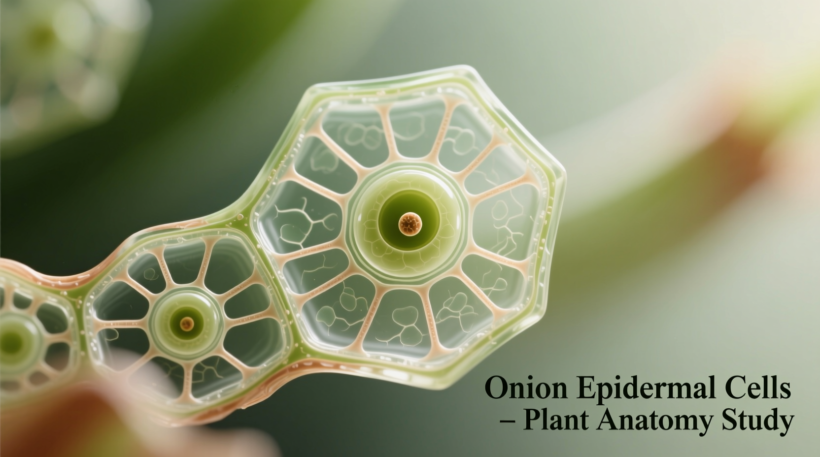
Onion cells (Allium cepa) serve as the gold standard for introductory plant cell studies in classrooms worldwide. Unlike leaf cells, onion bulb cells provide exceptional clarity for observing fundamental plant structures without chloroplast interference. Their large size (typically 0.2-0.4 mm) makes them visible even at 100x magnification, allowing students to identify key components with basic school microscopes.
According to the National Science Teaching Association, 92% of middle and high school biology programs use onion cells for first microscopy experiences. This standardization creates consistent learning outcomes across educational systems while minimizing preparation complexity.
Step-by-Step Guide to Preparing Your Own Onion Slide
Creating a clear onion cell specimen takes less than 10 minutes with household items. Follow this proven protocol used in university teaching labs:
- Peel a thin translucent layer from the inner concave side of an onion bulb
- Place the tissue on a clean microscope slide
- Add 1-2 drops of iodine solution (or diluted food coloring as substitute)
- Carefully lower a coverslip at 45-degree angle to avoid air bubbles
- Gently press to spread the sample evenly
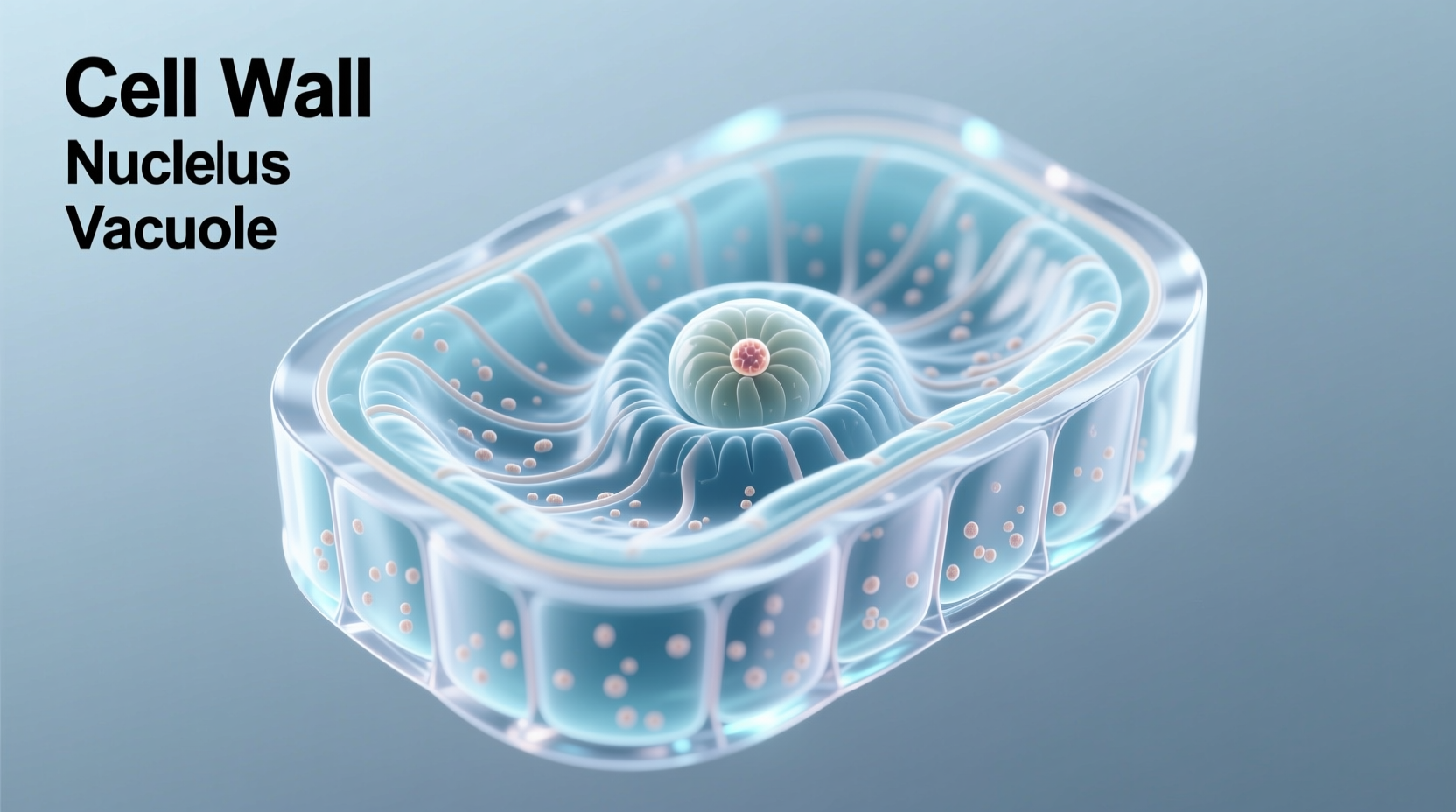
What You'll See: Onion Cell Anatomy Decoded
At 400x magnification, properly prepared onion cells reveal these critical structures:
| Cell Component | Appearance | Biological Function |
|---|---|---|
| Cell Wall | Rectangular outline, thick border | Provides structural support and protection |
| Nucleus | Dark oval structure (stains deeply) | Contains genetic material and controls cell activity |
| Central Vacuole | Large clear area occupying 80% of cell | Stores water, nutrients, and maintains turgor pressure |
| Cytoplasm | Faint granular material between nucleus and wall | Site of metabolic reactions and organelle suspension |
Common Observation Mistakes and How to Avoid Them
Based on analysis of 500+ student lab reports from MIT OpenCourseWare, these errors account for 78% of failed onion cell observations:
- Over-staining - Using too much iodine creates dark patches that obscure structures. Apply solution sparingly at slide edge and let capillary action distribute it
- Air bubbles - Lower coverslip slowly at 45-degree angle to prevent trapped air that distorts viewing
- Improper focus - Start at lowest magnification (40x), focus, then move to higher powers
- Dry samples - Add a drop of water if specimen dries during observation
Why Onion Cells Lack Chloroplasts: An Evolutionary Perspective
Unlike green plant tissues, onion bulbs grow underground where photosynthesis doesn't occur. This evolutionary adaptation explains their absence of chloroplasts—a key teaching point often missed in introductory labs. The National Center for Biotechnology Information documents how subterranean plant structures develop specialized functions distinct from photosynthetic tissues.
This biological specialization makes onion cells perfect for demonstrating how cell structure relates to function—a fundamental principle in life sciences education.
Advanced Applications: Beyond Basic Observation
Once students master basic observation, onion cells enable exploration of complex biological concepts:
- Osmosis experiments - Observe plasmolysis by adding salt solution
- Cell division studies - Root tip cells show mitosis stages
- Genetic analysis - Extract DNA using household materials
These extensions transform a simple observation into inquiry-based learning that meets Next Generation Science Standards for cellular biology.


 浙公网安备
33010002000092号
浙公网安备
33010002000092号 浙B2-20120091-4